

Modified Blood Oxygen Levels Detection
Written by M. Ananthi, T. Mangayarkarasi, R. K. Sai Suni Kumar, G. Jaya DarshiniI, and U. V. Kalaivani
A consumer healthcare device called an OXIMETER is used to measure blood oxygen levels. It is optimized for providing smart elderly care applications. The oximeter prototype in its present form, however, does not produce correct results in people with scleroderma, people with decreased tissue perfusion (as is the case in individuals with anemia), and people with digital ulcers. Scleroderma is an autoimmune illness in which the body attacks its own cells because it believes they are foreign invaders. This makes the skin rough, thus making it difficult to accurately measure blood oxygen levels. OXILASER is a gadget that combines an oximeter and a laser. The laser aids in penetrating through rough skin to easily and accurately measure blood oxygen levels.
Introduction
The pandemic has opened our eyes to a number of new possibilities, as many industries have become increasingly digitized. This digitization also occurred in the medical industry. Regular check-ups and in-person appointments between doctors and patients were hampered by COVID-19. In many instances, only COVID-19 wards were open, and many hospitals and clinics were closed. As a result, many patients turned to home monitoring and e-medicine. Scleroderma patients are a specific group of people that need their blood oxygen levels checked on a frequent basis. Scleroderma mostly affects certain tissues, some of which exist in the lungs. Fibrosis of these tissues in the lungs leads to Interstitial Lung Disease (ILD). ILD (fibrosis of lungs) causes shortness of breath. ILD and scleroderma frequently coexist. The frequency of fatalities rose as a result of closed clinics, as patients with ILD/scleroderma found it challenging to monitor their oxygen levels. Scleroderma (SCLERO-hard DERMA-skin) is an autoimmune disease where the immune system attacks the connective tissues, thereby generating dead cells in large numbers, which causes thickening of the skin. It also attacks blood vessels, internal organs, and the digestive tract. There is no cure for this disease, but the progression of the disease can be curbed, to an extent, through medications. Figure 1 illustrates the symptoms of scleroderma.
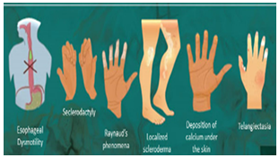
Figure 1: Oximeter for various elderly care applications.
Esophageal Dysmotility:
Esophageal Dysmotility is a condition where it is difficult for the patients to swallow food, resulting in regurgitation.
Sclerodactyly:
Sclerodactyly is a symptom of scleroderma in which the fingers swell and nails curl inward.
Calcinosis Cutis:
Calcinosis cutis is a condition where insoluble calcium particles deposit under the skin, forming calluses and making the skin rough and sore.
There are two forms of scleroderma:
- Localized Scleroderma
Localized scleroderma is one manifestation of the condition in which the disease does not attack the internal organs, and only causes thickening and hardening of skin. Waxy patches (morphea) are also seen on the skin.
Raynaud’s phenomenon is a condition in which blood flow to the fingers and toes is restricted, which results in the change of color of the skin, finger soreness, numbness, etc.
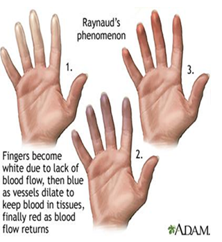
Figure 2: The color of the finger changes in scleroderma patients due to disrupted blood flow.
- Diffused Scleroderma:
Diffused scleroderma is one manifestation of the condition in which the disease affects the internal organs. The internal organs contain few connective tissues; hence the immune system attacks those tissues too, resulting in fibrosis. It affects the:- Gastro-Intestinal tract
- Kidneys
- Lungs
- Heart
Gastro – Intestinal Tract: In scleroderma patients, fibrosis of the intestine occurs, making the intestine weak. As a result, consumed food is not digested, resulting in bloating, abdominal pain, regurgitation, malabsorption, weight loss, etc.
Kidneys: Sclerosis renal crisis can be a life-threatening condition of scleroderma that occurs due to hypertension, which leads to renal failure.
Heart: Scleroderma causes scarring of tissues in the heart, which leads to a condition called PAH (Pulmonary Artery Hypertension), which affects the arteries present in the heart.
Interstitial Lung Disease(ILD)
One type of diffuse scleroderma is ILD. Lungs are formed of pseudostratified ciliated columnar epithelium, a kind of connective tissue. Because pulmonary fibrosis is an autoimmune illness, the immune system destroys the tissue in the lungs, leading to fibrosis. ILD makes breathing difficult. Blood oxygen levels need to be checked often in patients with ILD (Interstitial Lung Disease). Patients with scleroderma who are diagnosed with ILD early on can avoid fatalities.
An oximeter is a device used to measure blood oxygen levels. LEDs are typically used in oximeters to measure oxygen levels. These LEDs fail to penetrate through certain skin types. In scleroderma patients, the oximeter provided falsely low readings, and in most cases, the patient's finger's positioning even went unnoticed by the device. During the pandemic, it was necessary to monitor blood oxygen levels in all affected patients. Due to inaccurate measurements, the mortality rate among scleroderma patients rose. Oximeter readings fluctuate, even in healthy people. The oximeter's biggest flaw is its lack of precision. It showed figures that were between 5% and 9% higher or lower than true values.
It is necessary to accurately determine blood oxygen levels in order to reduce mortality. This is achieved by infusing LASER in an oximeter. The laser provides a narrow beam of light that can pass through any type of skin texture and improve the accuracy of the readings of blood oxygen levels.
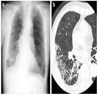
Figure 3: depicts the difference between lungs of a healthy human and that of lungs of a patient diagnosed with ILD (Interstitial Lung Disease), whose efficiency is much less compared to the lungs of a healthy human being.
Methodology
- Proposed solution
Oxilaser operates according to the photoplethysmography concept. Photoplethysmography is a technique for identifying changes in the microvascular blood of tissue's blood volume. The incident light is permitted to pass through the finger, and the photodiode detects the transmitted light. The blood absorbs the incident light, and the remaining light is sent to the photodiode. If the photodiode detects a lot of red light, it means that the body's blood is more oxygenated than usual, and if it detects a lot of IR LED, it means that the blood is more deoxygenated. - Working principle
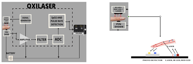
Figure 4:The system architecture of the device. Its components are explained below.
- INFRARED AND RED LEDS IN OXILASER: Infrared LED and RED LED are used in determining blood oxygen level. These lights are allowed to pass through the finger and are absorbed by the blood. The unabsorbed light is detected by the photodiode on the other end.
- LASER IN OXILASER: LASER (wavelength 632 nm - 650 nm) helps to penetrate through rough skin and detect blood oxygen levels in scleroderma patients. LASER is a monochromatic source of light, as it produces light of single wavelength.
- PHOTODIODE IN OXILASER: This plays a vital role, as the amount of oxygenated or deoxygenated blood levels are determined only by the amount of light detected by the photodiode.
- Red light>IR = More Oxygenated Blood
- Red light<IR =More Deoxygenated Blood
- SIGNAL GENERATOR: Signal generator is used to ensure accurate signal reading between humans and signal analyzer.
- AMPLIFIER: The amplifier converts signal from sensor to voltage.
- FILTER: Filter limits the signal (converted by amplifier) to a certain frequency.
- ANALOG TO DIGITAL CONVERTER (ADC): ADC converts analogue values signals to equivalent digital values.
- SPO2 AND PULSE RATE DETECTION: It is used as an indirect estimation of arterial oxygen saturation. This value is displayed in the LCD.
Testing and Results
A. Existing Systems
Existing systems for blood oxygen level detection include pulse oximetry, esophageal oximetry, forehead oximetry, and earlobe oximetry, all of which have difficulty accurately determining oxygen levels in scleroderma patients.
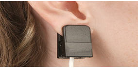
Figure 5
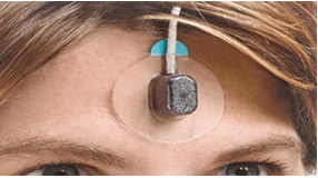
Figure 6
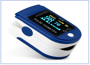
Figure 7
Figure 5, Figure 6, and Figure 7 depict earlobe oximetry, forehead oximetry, and pulse oximetry, respectively.
ABG TEST: An ABG test (Arterial Blood Gas Test) is performed to determine the blood's oxygen content. This laborious and time-consuming procedure yields exact results.
Under the direction of rheumatologist Dr. C. Balaji, a study of patients admitted to Sri Ramachandra Hospital in Chennai was conducted on 23rd June 2023. Dr. Balaji asserted that the Oxilaser could replace the oximeter in the near future due to its accuracy.
Dr. C. Balaji is a renowned rheumatologist who has nearly 17 years of experience. Rheumatologists diagnose and treat patients with autoimmune diseases.
B. Inferences
1. Comparison of blood oxygen levels in scleroderma patients versus healthy individuals
Oximeter values from 10 scleroderma patients were compared to those from 10 healthy individuals.
| S.No. | Blood oxygen levels in scleroderma patients | Blood oxygen level in healthy individuals |
| 1. | 66 | 99 |
| 2. | 42 | 99 |
| 3. | 53 | 98 |
| 4. | 90 | 99 |
| 5. | 32 | 97 |
| 6. | 76 | 92 |
| 7. | 0 | 96 |
| 8. | 34 | 99 |
| 9. | 85 | 98 |
| 10. | 59 | 94 |
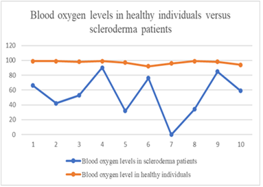
Figure 8 illustrates discrepancies in blood oxygen levels between scleroderma patients and healthy people. Here, the x axis is the no. of patients and y axis is the values of blood oxygen levels. We acquire nearly precise readings (as of the values prescribed by the doctor) in healthy individuals, whereas scleroderma patients receive artificially low numbers.
2. Comparison of blood oxygen levels in scleroderma patients using pulse oximetry versus ABG Test
| S.No. | Age | Gender | Blood oxygen levels in scleroderma patients using pulse oximetry | Blood oxygen level in scleroderma patients using ABG Test |
| 1. | 39 | F | 66 | 90 |
| 2. | 32 | F | 42 | 91 |
| 3. | 56 | F | 53 | 88 |
| 4. | 42 | F | 90 | 86 |
| 5. | 45 | F | 32 | 92 |
| 6. | 28 | M | 76 | 98 |
| 7. | 68 | M | 0 | 80 |
| 8. | 43 | F | 34 | 93 |
| 9. | 36 | M | 85 | 89 |
| 10. | 30 | F | 59 | 92 |
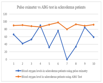
Figure 9 shows the differences between blood oxygen levels measured by pulse oximetry and those acquired by an ABG test in patients with scleroderma. The number of patients is on the x-axis and the blood oxygen level values are on the y-axis.
In the 7th patient, aged 68 years, blood oxygen levels were not obtained using the pulse oximeter, as it provided null values. In his ABG test, blood oxygen levels were measured to be 80. He was admitted into the ICU, as he was affected with ILD (Interstitial Lung Disease).
From the above table, a lot of variations can be observed between the two tests, which suggests the inefficiency of pulse oximeters.
3. Comparison of blood oxygen levels in healthy individuals using pulse oximetry versus ABG test
| S.No. | Blood oxygen levels in healthy individuals using pulse oximetry | Blood oxygen level in healthy individuals using ABG Test |
| 1. | 99 | 97 |
| 2. | 99 | 99 |
| 3. | 98 | 99 |
| 4. | 99 | 94 |
| 5. | 97 | 99 |
| 6. | 92 | 96 |
| 7. | 96 | 98 |
| 8. | 99 | 99 |
| 9. | 98 | 99 |
| 10. | 94 | 98 |
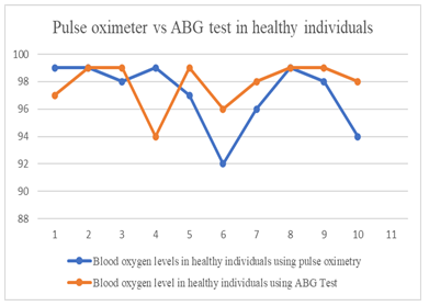
Figure 10: Blood oxygen levels in healthy people using a pulse oximeter are compared to those of an ABG test. Differences are evident, which suggests oximeters may show values that are 5% to 9% higher or lower than the actual readings.
C. Technical Information
Oxilaser produces combined values of heart sensor and MAX30100.
Heart Sensor:
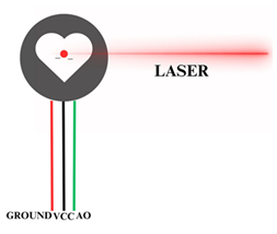
Figure 11
Heart sensor usually emits green LED, whereas in Oxilaser this LED is replaced with Red LASER. Figure 11 depicts the emission of LASER from Heart sensor.
OXILASER Circuit diagram:
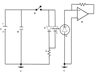
Figure 12: Depicts the circuit diagram of Oxilaser
The components pictured in Figure 12:
- A-BATTERY
- B-CAPACITOR
- C-GROUND
- D-SWITCH
- E-LED
- F-RESISTOR
- G-PHOTODIODE
- H-AMPLIFIER
- I-LASER DIODE
The LASER is emitted from the Heart sensor, and the IR LED and RED LED’s are emitted by MAX30102 as usual.
Heart sensor usually emits GREEN LED, whereas in Oxilaser it is replaced with LASER.
Laser:
The red LASER is used to penetrate through any skin texture. The wavelength of LASER used is between 632 nm - 650 nm (this wavelength is also being used in Low Level Laser Treatment (LLLT) to reduce pain in Rheumatoid Arthritis patients, and there are no reports of generation of carcinogenic cells in those patients). The photons released by LASER are absorbed by the mitochondria of the cell.
Conclusion and Future Scope
Scleroderma is an uncommon condition. Due to the inaccuracy of readings obtained by Oximeters, Interstitial Lung Disease was only diagnosed in late stages in scleroderma patients, which is the leading cause of mortality in patients with the disease. The use of Oxilaser lowers mortality and aids in the early diagnosis of ILD in people with scleroderma. Additionally, Oxilaser can be used to track how the COVID-19 virus affects COVID patients. The accuracy of this instrument will result in its widespread adoption, making it particularly suited for aged care in a smart city environment.
References
- Yashdeep Singh Pathania - Alternatives for erroneous finger probe pulse oximetry in systemic sclerosis patients during COVID-19 pandemic,2021, DOI: 10.1007/s00296-021-05032-w
- Fahad Gul, Amna Siddiqui, Prakhyath Srikaram , Nabeela Fatima - Scleroderma and interstitial lung disease - A case report,2022,DOI: 10.1016/j.amsu.2022.104143
- Marlies S Wijsenbeek, Catharina C Moor, Kerri A Johannson, Peter D Jackson, Yet H Khor , Yasuhiro Kondoh, Sujeet K Rajan, Gabriela C Tabaj, Brenda E Varela , Pieter van der Wal, Richard N van Zyl-Smit MD, Michael Kreuter, Toby M Maher - Home monitoring in interstitial lung diseases,2023, DOI:10.1016/S2213-2600(22)00228-4
- Kwang-Suk Seo, M.D., Jung Soo Kim, M.S., Wonsik Ahn, M.D., Kwang Suk Park, Ph.D., Hyun-Jeong Kim, M.D., Kwang-Won Yum, M.D., and Eui-Kyoung Goo, M.D - Estimation of Continuous Blood Pressure with Amplitude of Photoplethysmogram and Pulse Transit Time of Finger and Toe,2022, DOI:10.4097/kjae.2007.53.2.159
- Ivan Corazza, Laura Cercenelli - Technologies for Hemodynamic Measurements: Past, Present and Future, 2022, DOI: 10.1016/B978-0-12-816861-5.00022-8.
- Geeta S Agashe 1, Joseph Coakley, Paul D Mannheimer - Forehead pulse oximetry: Headband use helps alleviate false low readings likely related to venous pulsation artifact,2022, DOI:10.1097/00000542-200612000-00010.
- Amaka Odonwodo; Talel Badri; Anis Hariz.- Scleroderma,2022
- Jens Klotsche, Kathryn S Torok, Ozgur Kasapcopur, Amra Adrovic,Maria Teresa Terreri - Application and performance of disease activity indices proposed for patients with systemic sclerosis in an international cohort of patients with juvenile systemic sclerosis,2023, DOI:10.1177/23971983231164700
- H. Chung, Christopher M. Walker, Stephen Hobbs - Imaging Features of Systemic Sclerosis-Associated Interstitial Lung Disease,2021, DOI: 10.3791/60300
- Khan F, Howard L, Hearson G, et al. Clinical utility of home versus hospital spirometry in fibrotic ILD: evaluation following INJUSTIS interim analysis. Ann Am Thorac Soc. 2022; 19: 506–09.
- Moor CC, van Leuven SI, Wijsenbeek MS, Vonk MC. Feasibility of online home spirometry in systemic sclerosis-associated interstitial lung disease: a pilot study. Rheumatology 2021; 60: 2467–71.
- Johannson KA, Lethebe BC, Assayag D, et al. Travel distance to subspecialty clinic and outcomes in patients with fibrotic interstitial lung disease. Ann Am Thorac Soc 2022; 19: 20–27.
- Moor CC, van den Berg CAL, Visser LS, Aerts J, Cottin V, Wijsenbeek MS. Diurnal variation in forced vital capacity in patients with fibrotic interstitial lung disease using home spirometry. ERJ Open Res 2020; 6: 00054-2020.
- Noth I, Cottin V, Chaudhuri N, et al. Home spirometry in patients with idiopathic pulmonary fibrosis: data from the INMARK trial. Eur Respir J 2021; 58: 2001518.
- Wijsenbeek MS, Bendstrup E, Valenzuela C, et al. Disease behavior during the peri-diagnostic period in patients with suspected interstitial lung disease: the STARLINER study. Adv Ther 2021; 38: 4040–56.
- Hoffmann-Vold AM, Allanore Y, Bendstrup E, et al. The need for a holistic approach for SSc-ILD—achievements and ambiguity in a devastating disease. Respir Res 2020; 21: 197.
- Avitzur N, Noth EM, Lamidi M, et al. Relative environmental and social disadvantage in patients with idiopathic pulmonary fibrosis. Thorax 2021; published online Dec 23. https://doi.org.10.1136/ thoraxjnl-2021-217652.
- Haleem A, Javaid M, Singh RP, Suman R. Telemedicine for healthcare: capabilities, features, barriers, and applications. Sens Int 2021; 2: 100117.
- Nakshbandi G, Moor CC, Johannson KA, Maher TM, Kreuter M, Wijsenbeek MS. Worldwide experiences and opinions of healthcare providers on eHealth for patients with interstitial lung diseases in the COVID-19 era. ERJ Open Res 2021; 7: 00405-2021.
- Goobie GC, Nouraie M, Zhang Y, et al. Air pollution and interstitial lung diseases: defining epigenetic effects. Am J Respir Crit Care Med 2020; 202: 1217–24
- Sheikh A, Anderson M, Albala S, et al. Health information technology and digital innovation for national learning health and care systems. Lancet Digit Health 2021; 3: e383–96
- Ferini-Strambi, Luigi, and Maria Salsone. "COVID-19 and neurological disorders: are neurodegenerative or neuroimmunological diseases more vulnerable?" Journal of neurology (2020): 1-11.
- Peach E, Rutter M, Lanyon P, et al. Risk of death among people with rare autoimmune diseases compared with the general population in England during the 2020 COVID-19 pandemic. Rheumatology 2021; 60: 1902–09.
- Gupta L, Kharbanda R, Agarwal V, Misra DP, Agarwal V. Patient perspectives on the effect of the SARS-CoV-2 pandemic on patients with systemic sclerosis: an international patient survey. J Clin Rheumatol 2021; 27: 31–33.
- Sampaio-Barros PD, Medeiros-Ribeiro AC, Luppino-Assad AP, et al. SARS-CoV-2 vaccine in patients with systemic sclerosis:impact of disease subtype and therapy. Rheumatology (Oxford) 2022; 61: SI169–74.
- Hoffmann-Vold AM, Brunborg C, Tirelli F, et al. POS0054 The impact and outcome of COVID-19 on systemic sclerosis patients from the European scleroderma trial and research group (EUSTAR). Ann Rheum Dis 2021; 80 (suppl 1): 232–33.
- Nalbandian A, Sehgal K, Gupta A, et al. Post-acute COVID-19 syndrome. Nat Med 2021; 27: 601–15.
- Moiseev S, Avdeev S, Brovko M, et al. Rheumatic diseases in intensive care unit patients with COVID-19. Ann Rheum Dis 2021; 80: e16.
- S. Seifi, A. Khatony, G. Moradi, A. Abdi, and F. Najafi, “Accuracy of pulse oximetry in detection of oxygen saturation in patients admitted to the intensive care unit of heart surgery: Comparison of finger, toe, forehead and earlobe probes,” BMC Nursing, vol. 17, no. 1, pp. 1–7, Dec. 2018, doi: 010.1186/s12912-018-0283-1
- Maddipatla et al., “A polyimide-based force sensor fabricated using additive screen-printing process for flexible electronics,” In IEEE Access, vol. 8, pp. 207813-207821, doi: 10.1109/ACCESS.2020.3037703, 2020.
- Kang Zhang, Xiaohong Liu, Jun Shen, Zhihuan Li, Ye Sang, Xingwang Wu, Yunfei Zha, Wenhua Liang, Chengdi Wang, Ke Wang, Linsen Ye, Ming Gao, Zhongguo Zhou, Liang Li, Jin Wang, Zehong Yang, Huimin Cai, Jie Xu, Lei Yang, Wenjia Cai, Wenqin Xu, Shaoxu Wu, Wei Zhang, Shanping Jiang, Lianghong Zheng, Xuan Zhang, Li Wang, Liu Lu, Jiaming Li, Haiping Yin, Winston Wang, Oulan Li, Charlotte Zhang, Liang Liang, Tao Wu, Ruiyun Deng,Kang Wei, Yong Zhou, Ting Chen, Johnson Yiu-Nam Lau, Manson Fok, Jianxing He, Tianxin Lin, Weimin Li, and Guangyu Wang, “Clinically Applicable AI System for Accurate Diagnosis, Quantitative Measurements, and Prognosis of COVID-19 Pneumonia Using Computed Tomography ,” Cell 181, 1423–1433 June 11, 2020 Elsevier Inc., doi:10.1016/j.cell.2020.04.045
- Hughes M, Pauling JD, Moore A, Jones J. Impact of COVID-19 on clinical care and lived experience of systemic sclerosis: an international survey from EURORDIS-Rare Diseases Europe. J Scleroderma Relat Disord 2021; 6: 133–38.
- Denton CP, Campochiaro C, Bruni C, Distler O, Iagnocco A, Matucci Cerinic M. COVID-19 and systemic sclerosis: rising to the challenge of a pandemic. J Scleroderma Relat Disord 2021; 6: 58–65.
- Avouac J, Airó P, Carlier N, Matucci-Cerinic M, Allanore Y. Severe COVID-19-associated pneumonia in 3 patients with systemic sclerosis treated with rituximab. Ann Rheum Dis 2021; 80: e37.
- Strangfeld A, Schäfer M, Gianfrancesco MA, et al. Factors associated with COVID-19-related death in people with rheumatic diseases: results from the COVID-19 Global Rheumatology Alliance physician-reported registry. Ann Rheum Dis 2021; 80: 930–42.
- Adams HJA, Kwee TC, Yakar D, Hope MD, Kwee RM. Systematic review and meta-analysis on the value of chest ct in the diagnosis of coronavirus disease (COVID-19): Sol Scientiae, Illustra Nos. AJR Am J Roentgenol 2020; 215: 1342–50.
- Orlandi M, Landini N, Sambataro G, et al. The role of chest CT in deciphering interstitial lung involvement: systemic sclerosis versus COVID-19. Rheumatology 2022; 61: 1600–09.
- Matucci-Cerinic M, Hughes M, Taliani G, Kahaleh B. Similarities between COVID-19 and systemic sclerosis early vasculopathy: A “viral” challenge for future research in scleroderma. Autoimmun Rev 2021; 20: 102899.
- Palacios R, Patiño EG, de Oliveira Piorelli R, et al. Double-blind, randomized, placebo-controlled phase III clinical trial to evaluate the efficacy and safety of treating healthcare professionals with the adsorbed COVID-19 (Inactivated) vaccine manufactured by sinovac - PROFISCOV: a structured summary of a study protocol for a randomized controlled trial. Trials 2020; 21: 853.
- Schwarzkopf S, Krawczyk A, Knop D, et al. Cellular immunity in COVID-19 convalescents with PCR-confirmed infection but with undetectable SARS-CoV-2-specific IgG. Emerg Infect Dis 2021; 27: 122–2
Keywords: Oximeter, Oxilaser, Scleroderma, autoimmune disease, ILD (Interstitial Lung Disease), Photoplethysmography
This article was edited by Bernard Fong.
To view all articles in this issue, please go to August 2023 eNewsletter. For a downloadable copy, please visit the IEEE Smart Cities Resource Center.

To have the eNewsletter delivered monthly to your inbox, join the IEEE Smart Cities Community.
Past Issues
To view archived articles, and issues, which deliver rich insight into the forces shaping the future of the smart cities. Older eNewsletter can be found here. To download full issues, visit the publications section of the IEEE Smart Cities Resource Center.
IEEE Smart Cities Newsletter Editors

Managing Editor

Assistant Managing Editor
IEEE Smart Cities Publications Editorial Board
Luis M. Fernandez-Ramirez
Bernard Fong
Sergii Kushch
Prasad Enjeti
Rabie Ramadan
Ramkumar Lakshminarayanan
Francesco Flammini
Fateh Krim
Maria Pia Fanti
Kristina Kunert
Vladimir Orlic
Asif Ali Ahmed R
Werbeston Oliveira
Emilio Ghiani
Shafi Khadem
Nikumani Choudhury
Mohammad Zeyad
S.M. Masum Ahmed
Payman Dehghanian
Sona N Thadevus
Kumaresan Natarajan
Lues Felipe Gaitan Cubides
Chao Shen
Balaji Sankarshanan
Chun Sing Lai
Zhang Wei
Shashikant Patil
Sai Munikoti
Akhil Jabbar Meerja
Click here for more info
Loi Lei Lai
Editor-in-Chief
IEEE Smart Cities eNewsletter